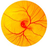
Many elaborate physiological processes take place during the transformation of the embryo from egg to chick. These processes are respiration, excretion, nutrition, and protection.

For the embryo to develop without any anatomical connection to the hen's body, nature has provided membranes outside the embryo's body to enable the embryo to use all parts of the egg for growth and development. These "extra-embryonic" membranes are the:
- yolk sac
- amnion
- chorion
- allantois
The yolk sac is a layer of tissue growing over the surface of the yolk. Its walls are lined with a special tissue that digests and absorbs the yolk material to provide sustenance for the embryo. Yolk material does not pass through the yolk stalk to the embryo even though a narrow opening in the stalk is still in evidence at the end of the incubation period. As embryonic development continues, the yolk sac is engulfed within the embryo and is completely reabsorbed at hatching. At this time, enough nutritive material remains to adequately maintain the chick for up to two days.
The amnion is a transparent sac filled with a colorless fluid that serves as a protective cushion during embryonic development. This amniotic fluid also permits the developing embryo to exercise. The embryo is free to change its shape and position while the amniotic fluid equalizes the external pressure. Specialized muscles also develop in the amnion, which by smooth, rhythmic contractions gently agitate the amniotic fluid. The slow and gentle rocking movement apparently aids in keeping the growing parts free from one another, thereby preventing adhesions and resultant malformations.
The chorion serves as a contain for both the amnion and yolk sac. Initially, the chorion has no apparent function but later the allantois fuses with it to form the choric-allantoic membrane. This brings the capillaries of the allantois into direct contact with the shell membrane, allowing calcium reabsorption from the shell.
The allantois has four functions:
- It serves as an embryonic respiratory organ.
- It receives the excretions of the embryonic kidneys.
- It absorbs albumen, which serves as nutriment (protein) for the embryo.
- It absorbs calcium from the shell for the structural needs of the embryo.
The allantois differs from the amnion and chorion in that it arises within the body of the embryo. In fact, its proximal portion remains intra-embryonic throughout the development.
Functions of the Embryonic Membranes
Special temporary organs or embryonic membranes are formed within the egg, both to protect the embryo and to provide for its nutrition, respiration and excretion. These organs include the yolk sac, amnion and allantois.
The yolk sac supplies food material to the embryo. The amnion, by enclosing the embryo, provides protection. The allantois serves as a respiratory organ and as a reservoir for the excreta. These temporary organs function within the egg until the time of hatching and form no part of the fully developed chick.
Functions of the Embryonic Blood Vessels
During the incubation period of the chick, there are two sets of embryonic blood vessels. One set, the viteline vessels, is concerned with carrying the yolk materials to the growing embryo. The other set, the allantoic vessels, is chiefly concerned with respiration and with carrying waste products from the embryo to the allantois. When the chick is hatched, these embryonic blood vessels cease to function.
Notice the various embryonic membranes in this picture of a nine day embryo.
- The clear fluid surrounding the chick is the amnion.
- The yellow area covered with a blood system is the yolk sac.
- The dense blood system in the piece of egg shell is the allantois.
- The milky, clear material to the right of the shell is remaining white or albumen.
Pennsylvania State 4-H Office
- Email pa4h@psu.edu
- Office 814-863-3824
Pennsylvania State 4-H Office
- Email pa4h@psu.edu
- Office 814-863-3824


